CT has been saving lives since the early 1970s, but the detector principle used in most clinical scanners stayed remarkably stable for about half a century: convert X-rays into light, convert light into charge, then integrate the total signal. Photon-counting CT changes that foundation. Instead of weighing the total deposited energy in a detector element, it counts individual X-ray photons and sorts them by energy.
Why this matters
Photon-counting CT is not just another reconstruction algorithm. It changes the front end of the scanner. When the detector removes electronic noise and keeps energy information at acquisition, the image pipeline gains options that conventional energy-integrating CT cannot fully recreate later in software.
1. The 50-Year Detector Blind Spot
In a conventional energy-integrating detector (EID), X-ray photons exiting the patient strike a scintillator. The scintillator converts X-ray energy into visible light, and a photodiode converts that light into an electrical signal. The measured value is the integrated energy collected over a short time interval.
That system works extremely well. It made modern CT possible. But it also loses information. The detector does not know whether the signal came from a few higher-energy photons or many lower-energy photons. It also gives higher-energy photons more weight because the final signal is proportional to total deposited energy.
- Electronic noise remains in the signal: noise from conversion and readout is added before reconstruction.
- Low-energy photons are underweighted: these photons often carry useful contrast information, especially around iodine enhancement.
- Spectral information is mostly discarded: unless special dual-energy acquisition is used, a conventional detector cannot retrospectively separate photon energies.
- Spatial resolution is constrained: smaller detector pixels collect fewer photons, making the signal more vulnerable to electronic noise.
2. Direct Conversion: The Semiconductor Idea
Photon-counting CT removes the scintillator from the main detection path. A semiconductor sensor such as cadmium telluride (CdTe) or cadmium zinc telluride (CZT) directly converts incoming X-ray photons into electron-hole pairs. The amount of charge is proportional to the photon's deposited energy.
Under an applied bias voltage, that charge cloud drifts toward pixel electrodes. The readout ASIC sees the event as a fast voltage pulse. If the pulse height exceeds a chosen threshold, it is counted. If it is below the threshold, it is rejected as noise or low-energy contamination. With several thresholds, the system can sort photons into energy bins.
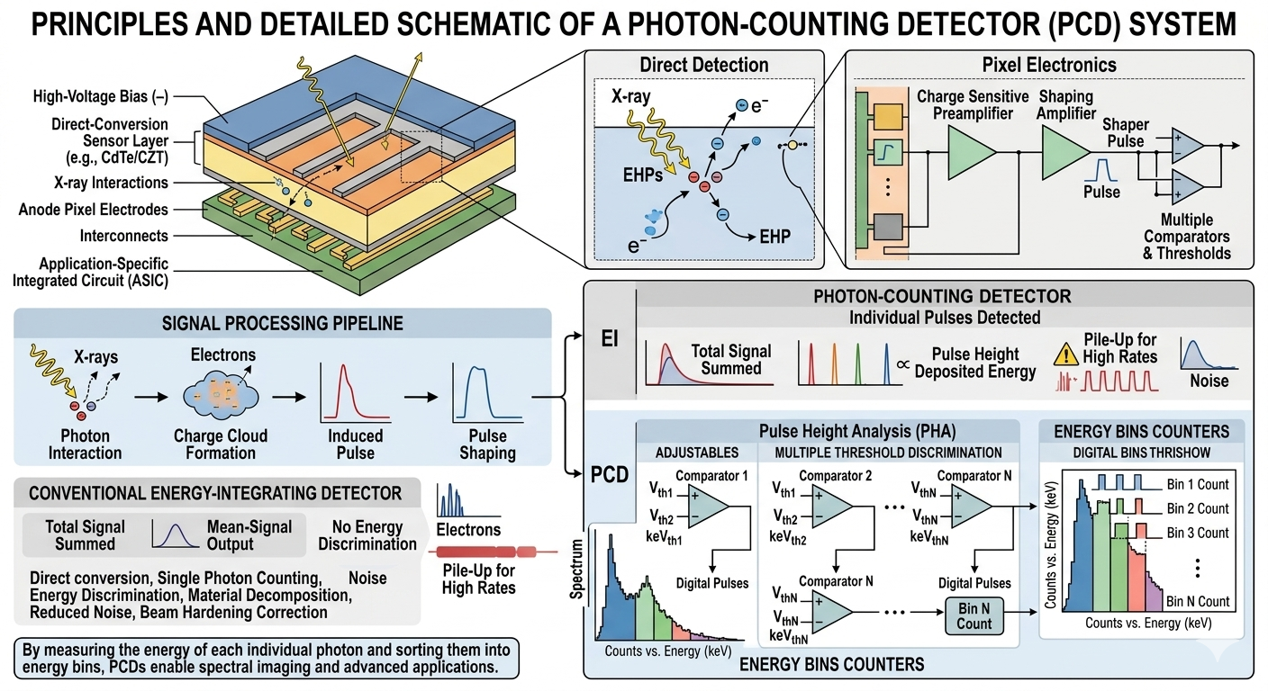
What changes at the detector?
Conventional CT measures the sum of energy arriving during a sampling window. Photon-counting CT treats each photon as an event. A direct-conversion semiconductor creates a charge pulse, the ASIC electronics compare that pulse against energy thresholds, and the scanner reconstructs counts across multiple energy bins.
An X-ray photon deposits energy inside CdTe, CZT, or a silicon detector geometry.
Electron-hole pairs are created, with charge magnitude linked to photon energy.
The ASIC compares pulse height with energy thresholds and rejects electronic noise.
Counts from multiple bins support VMI, iodine maps, Z-effective, and density images.
3. Materials: CdTe, CZT, and Silicon
Detector material selection is a major engineering decision. Clinical CT uses photon energies roughly in the diagnostic X-ray range, so the detector needs high stopping power, fast charge collection, stable room-temperature operation, and manufacturable crystal quality across a large detector area.
| Material | Why it is used | Engineering trade-off |
|---|---|---|
| Cadmium telluride (CdTe) | High atomic number and density give strong X-ray stopping power in a compact sensor. Siemens' cleared NAEOTOM Alpha uses CdTe photon-counting detectors. | Crystal growth, charge trapping, polarization, and uniformity are difficult at clinical detector scale. |
| Cadmium zinc telluride (CZT) | Closely related to CdTe and widely studied for energy-resolving X-ray and gamma-ray detection. | Material defects, yield, cost, and charge transport consistency affect spectral accuracy. |
| Silicon | Excellent electronics ecosystem, mature fabrication, and useful performance in some edge-on strip detector designs. | Lower atomic number means lower stopping efficiency unless geometry is extended to increase path length. |
4. Energy Thresholds and Spectral Data
The simplest photon-counting detector could use one threshold: count any pulse above a minimum energy and reject the rest. Clinical CT becomes more powerful when several thresholds are used at the same time. A photon that crosses a lower threshold but not a higher one falls into a lower energy bin; a photon crossing multiple thresholds is assigned according to the detector's binning logic.
This is the core reason photon-counting CT is often called spectral CT. The scanner does not need to run two separate tube voltages or add a second scan just to obtain energy-dependent information. The energy discrimination is built into the detector electronics.
Virtual monoenergetic images
Energy-bin data can synthesize images as if they were acquired with a narrower photon energy. Low-keV images often improve iodine conspicuity; higher-keV images can reduce some artifacts.
Material decomposition
Because attenuation changes with photon energy, spectral data can help separate basis materials such as iodine, calcium, soft tissue, and water-like components.
5. PCD-CT vs Conventional EID-CT
The difference is not only sharper pictures. The detector physics changes noise behavior, contrast weighting, dose efficiency, and what can be reconstructed from one acquisition.
| Feature | Energy-integrating CT | Photon-counting CT |
|---|---|---|
| Detection | Scintillator converts X-rays to light; photodiode integrates total signal. | Semiconductor directly converts each photon event into charge. |
| Noise handling | Electronic noise contributes to the measured signal. | Low pulse-height thresholds can reject electronic noise before image formation. |
| Energy information | Mostly lost unless dual-energy methods are used. | Captured intrinsically through multiple energy thresholds. |
| Spatial resolution | Smaller pixels eventually suffer from signal and noise limits. | Smaller effective pixels become more practical because electronic noise is suppressed. |
| Clinical workflow | Well-established, robust, and widely available. | Powerful but still premium, protocol-dependent, and less widely deployed. |
6. The NAEOTOM Alpha and Clinical Translation
The first clinically cleared photon-counting CT system was Siemens Healthineers' NAEOTOM Alpha. The FDA 510(k) letter is dated September 30, 2021 and identifies it as a computed tomography X-ray system. In the 510(k) summary, the device is described as a dual-source CT scanner with two photon-counting detectors based on cadmium telluride, using photon-counting data for reconstruction.
The FDA document also notes that images can support diagnosis, treatment preparation, radiation therapy planning, and low-dose lung cancer screening in high-risk populations. That matters for students because it shows photon counting was not cleared as a laboratory trick. It entered clinical CT as a full diagnostic platform with workflow, post-processing, and planning applications.
The market has also moved since the first 2021 clearance. Siemens Healthineers received FDA clearance for an expanded NAEOTOM Alpha class in 2025, and GE HealthCare announced FDA 510(k) clearance for its Photonova Spectra photon-counting CT in March 2026. So students should now see photon-counting CT as an emerging multi-vendor clinical category, not only a single flagship scanner.
Balanced interpretation
Photon-counting CT is a major detector advance, but it does not automatically make every scan superior. Protocol choice, patient size, dose settings, reconstruction method, task type, contrast timing, and radiologist experience still decide whether the theoretical detector advantage becomes a clinical advantage.
7. Why It Took So Long to Reach the Clinic
The idea of counting radiation events is not new. Nuclear medicine, particle physics, and laboratory detectors have used event-counting ideas for decades. CT was harder because the detector must work under very high X-ray flux, across a large field of view, while the gantry rotates, and while the system generates images fast enough for clinical workflow.
Early photon-counting CT research was easier in small-animal scanners because flux and field size were lower. Whole-body human CT created a different scale of problem: many more detector channels, much faster count rates, high manufacturing consistency, tight thermal control, and robust correction algorithms. This is why the field had promising papers long before hospitals could buy a clinical scanner.
- 1990s and early 2000s: semiconductor detectors matured in nuclear medicine, X-ray spectroscopy, and preclinical CT work.
- Prototype era: research systems showed high-resolution and multi-energy potential, but the clinical count-rate problem remained difficult.
- Clinical prototype era: academic centers tested whole-body photon-counting CT prototypes, building evidence for spatial resolution, noise behavior, and spectral use.
- 2021: FDA clearance of NAEOTOM Alpha turned photon-counting CT from a research platform into a clinical product category.
- 2025-2026: expanded FDA-cleared product families and new vendor clearances signalled a broader multi-vendor market.
8. System Specifications: What the Numbers Mean
Specification sheets can look like marketing unless you connect each number to an imaging consequence. For photon-counting CT, the most important numbers usually relate to detector material, pixel size, z-coverage, temporal resolution, thresholds, reconstruction modes, and whether the system uses one or two source-detector chains.
| Specification | Why a physicist cares | Why an engineer cares |
|---|---|---|
| Detector pixel pitch | Smaller effective pixels improve high-contrast spatial resolution and reduce partial-volume effects. | Smaller pixels increase channel count, heat, data throughput, and correction complexity. |
| Energy thresholds | Threshold placement affects noise rejection, material separation, iodine contrast, and quantitative accuracy. | Threshold circuits must remain stable across temperature, time, and detector channel variation. |
| Temporal resolution | Important for cardiac CT, motion artifact, and coronary artery assessment. | Depends on gantry rotation, source geometry, synchronization, and reconstruction pipeline performance. |
| Ultra-high-resolution mode | Useful for lung, bone, temporal bone, stents, and small structures. | May use different detector readout, z-coverage, dose settings, and reconstruction demands. |
| Spectral reconstruction | Enables VMI, iodine maps, effective atomic number, and electron density estimation. | Requires calibrated energy response, correction for pile-up and charge sharing, and stable DICOM output workflows. |
9. Technical Problems Engineers Had to Solve
Counting individual photons sounds simple until you place the detector in a clinical CT scanner. The photon flux is enormous, the detector rotates rapidly, and the system must operate with medical-device reliability. Several problems explain why the idea took decades to become a commercial whole-body CT system.
Pulse pile-up
If two photons arrive so close together that their charge pulses overlap, the electronics may count them incorrectly or interpret them as a single higher-energy event. This is called pile-up. It becomes more important at high flux, such as thick body regions or high-output protocols.
Charge sharing
A photon absorbed near a pixel boundary can create a charge cloud that spreads into neighboring pixels. One real photon may then look like two smaller events. This corrupts count accuracy and spectral accuracy unless anti-coincidence logic or correction algorithms are used.
K-escape and fluorescence effects
In high-Z semiconductor materials, characteristic X-rays can be produced and escape the original interaction site. The recorded energy may be lower than the incoming photon energy, or energy may be deposited in a neighboring pixel. This is another reason spectral calibration is a serious engineering task.
Detector uniformity and calibration
A CT detector is not one pixel. It is a large, curved array of many detector elements, readout channels, thresholds, gains, and temperature-sensitive electronics. Each channel must behave consistently enough that reconstruction algorithms can trust the raw data.
10. Calibration, QA, and the Medical Physics View
Photon-counting CT creates new quality assurance questions. A conventional CT QA program already checks CT number accuracy, uniformity, slice thickness, noise, resolution, dose output, and artifact behavior. Photon-counting CT adds energy-dependent behavior on top of that. The detector is no longer only asking, "How much signal arrived?" It is asking, "How many photons arrived in each energy range?"
That means spectral calibration becomes part of image quality. If thresholds drift, if one detector region bins photons differently from another, or if pile-up correction is imperfect, quantitative spectral outputs can shift. For routine visual diagnosis this may be subtle; for iodine quantification, electron density estimation, or longitudinal oncology measurements, it matters more.
Task-based image quality
A physicist will ask whether photon counting improves the specific diagnostic task: small vessel visibility, low-contrast lesion detection, stent lumen assessment, lung nodule margin reading, or material separation.
Dose optimization
The detector may allow lower dose for some tasks, but dose reduction must be proven against the clinical question. The goal is not the lowest dose; it is the right image quality at justified dose.
11. How Different Clinical Teams See PCD-CT
A good technology guide should not describe the machine from only one viewpoint. Radiologists, physicists, engineers, radiographers, and service teams notice different things.
Radiologist view
Sharper small structures, improved iodine contrast, reduced blooming in calcified vessels, better temporal bone detail, and more routine access to spectral reconstructions.
Medical physicist view
Dose efficiency, task-based image quality, spectral accuracy, CT number stability, protocol validation, quality control, and how reconstruction affects quantitative data.
Engineer view
Detector material behavior, ASIC heat, threshold calibration, gantry data throughput, service diagnostics, replacement cost, and long-term reliability.
Radiographer view
Patient setup, protocol selection, contrast timing, scan speed, artifact handling, post-processing workflow, and avoiding overcomplicated spectral outputs for routine studies.
12. Clinical Applications
Cardiac and vascular imaging
Cardiac CT benefits from high spatial resolution, reduced blooming artifact around calcification and stents, and the option to generate virtual monoenergetic images. In vascular imaging, spectral data can improve iodine conspicuity and may allow lower contrast volumes in selected protocols.
Calcified plaque is a good example of why spatial resolution and spectral behavior both matter. On conventional CT, dense calcium can appear larger than it is because of blooming, obscuring the vessel lumen. Photon-counting CT can reduce the size of that blur through smaller detector elements and sharper kernels, while spectral reconstructions may help separate calcium from iodine-enhanced blood.
Lung and thoracic imaging
Fine lung structures, small airways, fissures, and nodule margins are natural targets for high-resolution CT. Photon-counting detectors can make ultra-high-resolution acquisitions more practical, although dose, motion, reconstruction kernel, and clinical question still matter.
In thoracic imaging, the clinical value is not only prettier images. Better visualization of airway walls, emphysema patterns, interstitial lung disease detail, and small nodules can affect follow-up decisions. But interpretation must remain disciplined: higher resolution can reveal more findings, and more findings are not always more clinically useful unless they change management.
Musculoskeletal and temporal bone imaging
Small bone structures, trabecular detail, erosions, implant interfaces, and temporal bone anatomy can benefit from improved spatial resolution. This is one of the clearer cases where smaller detector pixels translate into visible detail.
Temporal bone CT is a useful student example because the target anatomy is tiny: ossicles, semicircular canals, cochlear structures, and bony canals. If the detector can preserve high spatial frequencies, the clinical reader can evaluate anatomy that may be blurred in a lower-resolution acquisition.
Oncology and contrast imaging
Spectral reconstructions can help quantify iodine uptake, reduce contrast dose in selected situations, and improve lesion conspicuity. Future K-edge contrast agents could extend this toward multi-material or molecular CT, but that is still an evolving area rather than routine practice.
Oncology is also where quantitative repeatability becomes important. If a tumor is being followed across treatment, the question may not simply be "is it visible?" but "has enhancement changed, has necrosis increased, has perfusion behavior shifted, or can a lesion be separated from surrounding tissue more reliably?" Photon-counting CT may support those questions, but validation must be protocol-specific.
Radiotherapy planning
Radiotherapy planning depends on accurate electron density information for dose calculation. Conventional planning CT uses HU-to-density calibration curves. Spectral CT and photon-counting CT may improve tissue characterization and electron density estimation, particularly where metal, contrast, or tissue composition complicates conventional calibration.
For radiotherapy physicists, this is one of the most interesting areas. Treatment planning systems convert image data into dose calculation inputs. If spectral CT can reduce uncertainty in electron density or improve tissue segmentation, it could influence planning accuracy. The practical question is whether the improvement survives the entire workflow: acquisition, reconstruction, contouring, density assignment, dose calculation, and adaptive review.
13. Data Pipeline: From Counts to Clinical Images
Photon-counting CT does not end at the detector. The raw measurement is a set of counts per detector pixel per projection per energy bin. Reconstruction software then converts those counts into images. Depending on the scanner and protocol, the output may include conventional CT images, ultra-high-resolution images, virtual monoenergetic images, iodine maps, calcium-suppressed views, virtual non-contrast images, effective atomic number maps, or electron density images.
This creates a new workflow question: which images should be sent to PACS by default, and which should remain available for post-processing? Too few outputs and the spectral value is hidden. Too many outputs and radiologists and radiographers face clutter. A mature photon-counting CT workflow is partly a human factors problem.
| Output | What it means | Where it can help |
|---|---|---|
| Conventional CT image | A familiar grayscale CT image reconstructed from the acquisition. | Routine reporting, comparison with prior scans, general workflow. |
| Virtual monoenergetic image | Image synthesized to approximate a chosen X-ray energy. | Iodine contrast enhancement, artifact reduction, vascular imaging. |
| Iodine map | Material-decomposition output estimating iodine distribution. | Contrast perfusion, lesion characterization, pulmonary embolism evaluation. |
| Electron density image | Quantitative output relevant to dose calculation and tissue modeling. | Radiotherapy planning and physics validation. |
14. What Students Should Not Misunderstand
- Photon counting does not remove radiation dose: CT still uses ionizing X-rays. The detector may use the dose more efficiently, but justification and optimization remain essential.
- Spectral data is not magic: material decomposition needs calibration, assumptions, and careful interpretation.
- Better hardware does not replace protocol design: scan mode, pitch, kV, mAs, kernel, contrast phase, and reconstruction still matter.
- Access is unequal: premium systems appear first in research hospitals and advanced imaging centers, while routine clinical access expands slowly.
15. Future Directions
The next phase is likely to involve wider multi-vendor competition, lower-cost detector manufacturing, better spectral calibration, AI-assisted reconstruction tuned for photon-counting noise statistics, and stronger integration with oncology and cardiovascular workflows.
The most interesting long-term direction is not only "sharper CT." It is quantitative CT: images that describe tissue composition more directly. Iodine concentration, effective atomic number, electron density, calcium characterization, and perhaps future K-edge contrast agents could make CT less like a grayscale shadow image and more like an energy-resolved tissue measurement.
K-edge contrast agents
A K-edge is a sharp jump in X-ray attenuation at a material-specific energy. If a contrast agent has a K-edge in the diagnostic CT energy range, a photon-counting detector can potentially identify that material more selectively. Iodine is already used clinically, but future research agents based on elements such as gadolinium, bismuth, gold, or nanoparticles could support multi-agent imaging. The clinical promise is attractive, but safety, pharmacology, regulatory approval, cost, and reconstruction validation are major barriers.
AI reconstruction and detector-aware models
Deep learning reconstruction is not unique to photon-counting CT, but PCD data has different noise and spectral structure from conventional CT. That means future models can be trained specifically for count statistics, energy-bin correlations, and material-decomposition outputs. The engineering risk is also clear: AI can make images look cleaner while hiding uncertainty if validation is weak.
Beyond diagnostic CT
Photon-counting ideas also appear in SPECT, PET detector logic, mammography research, X-ray spectroscopy, synchrotron imaging, and laboratory microscopy. The implementation changes by photon energy and count-rate requirement, but the core idea remains the same: detect events, preserve energy or timing information when useful, and avoid throwing away signal detail too early.
16. Study Summary
- Conventional CT detectors integrate total deposited energy after a scintillator and photodiode chain.
- Photon-counting detectors use direct semiconductor conversion to count individual photon events.
- Energy thresholds reject electronic noise and divide photons into spectral bins.
- CdTe and CZT offer high stopping power but bring crystal growth and charge-transport challenges.
- Key limitations include pulse pile-up, charge sharing, K-escape, calibration drift, cost, and clinical access.
- The first FDA-cleared clinical photon-counting CT system, NAEOTOM Alpha, received clearance on September 30, 2021, and the category has since expanded.
Student project idea
Simulate two detector models in Python: one energy-integrating detector that sums all photon energies with electronic noise, and one photon-counting detector that applies a threshold and bins photons by energy. Vary pixel size, count rate, and threshold level. Plot how signal-to-noise and material contrast change.
References and Further Reading
- FDA 510(k) clearance letter and summary for NAEOTOM Alpha, K211591
- Siemens Healthineers NAEOTOM Alpha product information
- Siemens Healthineers 2025 NAEOTOM Alpha class FDA clearance announcement
- GE HealthCare 2026 Photonova Spectra FDA clearance announcement
- The Technical Development of Photon-Counting Detector CT - PMC
- First Clinical Photon-counting Detector CT System: Technical Evaluation - PMC
- Clinical Applications of Photon-counting CT: Review of Pioneer Studies - PMC
- An Introduction to Photon-counting Detector CT for Radiologists - PMC